Abstract: Common and special sample processing methods and precautions.
Forward: The first step of successful experiment is learning to normalize the treatment of serum, plasma, and cell culture supernatants, cell lysate, tissue homogenates and other samples.
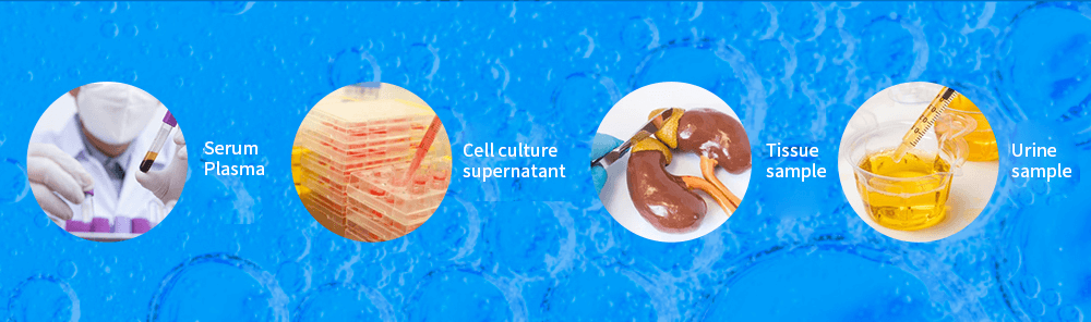
There are various types of samples used for ELISA experiments, including serum, plasma, cell culture supernatants, urine, and tissue homogenates. There are differences in pretreatment methods for different sample types. Proper sample pretreatment is the first step to ensure the accuracy of ELISA experiments. Here, there are several different sample types of processing methods will be introduced:
Common Sample Processing
1. Serum
Serum is the most commonly used sample for ELISA, and the pretreatment is also very simple.
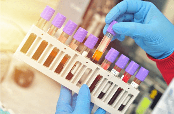
① Blood samples were collected using tubes without pyrogen and endotoxin or centrifuge tubes. Serum was separated by placing the test tubes or centrifuge tubes for 1 hour at room temperature or 2~8℃ overnight.
② The upper layer of serum was carefully collected by centrifugation for 20 min at 1000×g at 2~8℃.
2. Plasma
① Blood samples were collected using blood collection tubes containing anticoagulants or centrifuge tubes, and EDTA-Na2 was recommended as anticoagulant. Centrifuge samples for 15 min at 1000×g at 2~8℃ within 30 min of collection. Collect the supernatant to carry out the assay.
② Common anticoagulants include: EDTA-Na2, heparin sodium, sodium citrate, etc.
3. Cell Culture Supernatants
The cell culture supernatants were sucked into the centrifuge tube. Centrifuge samples for 15 min at 1000×g at 2~8℃, remove cell debris and impurities. Collect the supernatant to carry out the assay.
4. Cell Lysates
① The adherent cells were gently cleaned with precooled PBS, digested with trypsin, centrifuged for 5 min at 1000×g and then collected; Suspension cells can be collected directly by centrifugation.
② The collected cells were washed 3 times with cold PBS. In each 106 cells, 150~200 μL PBS was added for re-suspension (it is recommended to add protease inhibitors to PBS, if the content is very low, the volume of PBS can be reduced), and the cells were broken by repeated freeze-thaw or ultrasound;
③ The extraction solution was centrifuged for 10 min at 1500×g at 2~8℃, and the supernatant was detected.
5. Tissue Homogenates
① Rinse the tissue with pre-cooled PBS (0.01M, PH7.4) to remove the residual blood or impurities on the surface;
② Weigh the tissue block and cut it into pieces as small as possible so that it can be fully homogenized;
③ Add an appropriate amount of pre-cooled PBS (generally according to the weight to volume ratio of 1:9, such as 1g tissue sample corresponding to 9mL PBS, the specific volume can be adjusted according to the needs of the experiment, and make a record. It is recommended to add protease inhibitors to PBS) and fully homogenize in ice or ice bath with a glass homogenizer. In order to further lysis of tissue cells, the homogenate can be broken by ultrasound or frozen and thawed repeatedly.
④ The homogenate was sucked into the centrifuge tube, centrifuged for 5~10 min at 5000×g at 2~8℃. Collect the supernatant to carry out the assay.
Special Sample Processing Method
Saliva
Saliva was collected using a sterile EP tube and centrifuged for 10 min at 4000×g at 2~8℃ to remove impurities. Collect the supernatant to carry out the assay. Fresh saliva samples should be used.
Urine
Urine samples were collected in a sterile container and centrifuged for 20 min at 1000×g at 2~8℃ to remove impurities. Collect the supernatant to carry out the assay.
Feces
Feces samples were collected in a sterile container and PBS buffer was added to the feces at a ratio of 9 mL/g (0.01 M, pH=7.4; optionally add 0.05 M EDTA) and oscillate on ice for 15 min. Centrifuge it for 5~10 min at 5000×g at 2~8℃, and then collect the supernatant to carry out the assay.
Sample Precautions
1. Sample Collection
When collecting blood samples, hemolysis should be avoided as much as possible, and bacterial contamination should also be avoided to avoid false positive results.
Avoid using hyperlipemic samples. Lipids affect the homogeneity of samples and the binding of antigens and antibodies, resulting in a decrease in the accuracy of measurements.
2. Sample Storage
After sample collection and treatment, if the sample is assayed within 1 week, it can be stored in 2~8℃; and if it cannot be assayed in time, please pack it according to the amount of one time and freeze it in the environment of -20℃ (assayed within 1 month) or -80℃ (assayed within 3 months). Avoid repeated freeze-thaw cycles. Centrifuge again before assaying to remove any additional precipitates that may appear after storage.
3. Sample Dilution
The detection range of the kit is different from the concentration range of the sample. If the concentration of the substance to be measured in the sample is higher than the highest value of the standard, please make an appropriate dilution according to the actual situation (Suggest consulting literature and doing pre-experiments).
4. Sample Composition
Commercial cracking reagents are not recommended when handling cell extracts or tissue samples. Some components of cracking reagents may disrupt the spatial conformation of proteins, affect the binding of antigens and antibodies, or cause matrix interference, and ultimately affect the experimental results.