Cell Proliferation/Cytotoxicity/Viability
Cell proliferation detection is a basic experimental method to evaluate cell activity, genotoxicity and the effect of antitumor drugs. It generally reflects the growth status and activity of cells by analyzing the changes in the number of dividing cells. Cell activity is an important index to judge whether the cultured cells can grow normally under certain conditions in vitro. The detection of cell activity is an indirect method to detect cell proliferation ability. Cytotoxicity can be measured by changes in a number of indicators, including cell viability, cell proliferation, mitochondrial function, phospholipid deposition/lipid degeneration, DNA damage, and cell cycle.
Elabscience® provides cell proliferation and cytotoxicity/activity kits including EdU, CCK-8 and LDH. Depending on the type of sample and the purpose of your experiment, you can choose the appropriate kit according to your experimental requirements
EdU
- Simplicity: No antigen-antibody reaction is required, simple and efficient
- Sensitive: without antibody, the detection dye is only 1/500 of BrdU antibody, which is easy to spread and can be accurately detected even for a single proliferating cell
- Fast: No need to stay overnight, only 2~3 hours to complete the test
- Accurate: without DNA denaturation (acid hydrolysis, pyrolysis, enzymatic hydrolysis, etc.), it can effectively avoid sample damage caused by denaturation
- Compatible: almost no damage to the sample, easier to be labeled with a variety of antibodies or fluorescent proteins at the same time, can simultaneously detect other characteristics of the cell
| Product Name | Cat. No | Dye | Size |
|---|---|---|---|
| E-Click EdU Cell Proliferation Flow Cytometry Assay Kit (Green,FITC) | E-CK-A370 | FITC | 50 Assays/ 200 Assays |
| E-Click EdU Cell Proliferation Flow Cytometry Assay Kit (Green, Elab Fluor® 488) | E-CK-A371 | Elab Fluor® 488 | 50 Assays/ 200 Assays |
| E-Click EdU Cell Proliferation Flow Cytometry Assay Kit (Red, Elab Fluor® 647) | E-CK-A373 | Elab Fluor® 647 | 50 Assays/ 200 Assays |
| E-Click EdU Cell Proliferation Imaging Assay Kit (Green,FITC) | E-CK-A375 | FITC | 50 Assays/ 200 Assays |
| E-Click EdU Cell Proliferation Imaging Assay Kit (Green, Elab Fluor® 488) | E-CK-A376 | Elab Fluor® 488 | 50 Assays/ 200 Assays |
| E-Click EdU Cell Proliferation Imaging Assay Kit (Red, Elab Fluor® 594) | E-CK-A377 | Elab Fluor® 594 | 50 Assays/ 200 Assays |
| E-Click EdU Cell Proliferation Imaging Assay Kit (Red, Elab Fluor® 647) | E-CK-A378 | Elab Fluor® 647 | 50 Assays/ 200 Assays |
-
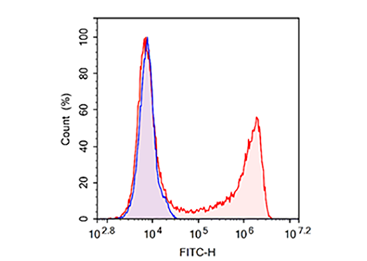 E-CK-A371
Jurkat cells were treated with 10 μM EdU for 4 h (red);without EdU (blue)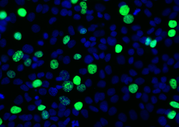 E-CK-A376
Hela cells were treated with 10 μM EdU for 2 h proliferate cells (green) and cells were counterstained with DAPI (blue)
Calcein AM/PI
Elabscience® Calcein AM/PI Double Staining Kit can be used to distinguish dead cells and living cells in mammals with esterase activity.
Calcein AM is the addition of acetyl methoxy methyl ester (AM) group to Calcein, which increases hydrophobicity and can easily penetrate the living cell membrane and enter the cell. Calcein AM itself has no fluorescence. After entering the cell, it is hydrolyzed by endogenous esterase in the cell to produce Calcein, a polar molecule with strong negative charge and cannot be retained in the cell through the cell membrane, while Calcein can emit strong green fluorescence ( Ex / Em = 494nm / 517nm ).Due to the lack of esterase, dead cells cannot or rarely produce Calcein, so only living cells are stained with strong green fluorescence, and dead cells cannot be stained or stained very weakly. The selective membrane permeability of dead cells is lost, and Propidium Iodide ( PI ) can enter the cell to specifically bind to doublestranded DNA and produce strong red fluorescence(Ex/Em = 535nm/617nm) to label dead cells. Therefore, the combination of Calcein AM and PI can perform double fluorescence staining on living cells and dead cells at the same time, which can be used for the detection of cell activity and cytotoxicity.
- Wide range of applications: Suitable for a variety of suspension cells and adherent cultured cells
- Diversification of detection:The results can be detected by flow cytometry and fluorescence microscopy
- Easy to operate:Doesn't need to spend time on reagent concentration fumbling, only takes about 15~30 min
- Low toxicity:Doesn't affect cell differentiation and proliferation
- Cost-effective:The reagent component is complete, and the buffer contains components to prevent Calcein's fluorescent spill
| Product Name | Cat. No | Size |
|---|---|---|
| Calcein AM/PI Double Staining Kit | E-CK-A354 | 100/500/2000 Assays |
| Calcein AM Solution(100 µM) | E-CK-A164 | 100 T/500 T/500 T*10 |
| PI Solution(750 µM) | E-CK-A165 | 100 T/500 T/500 T*10 |
| Calcein AM Assay Buffer | E-CK-A153 | 100 mL |
-
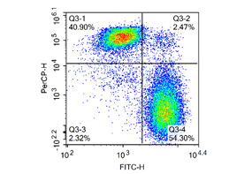 Figure1:Jurkat cells were placed at 4 °C for 20 days, then stained with Calcein-AM / PI Double Staining Kit and detected by flow cytometry.
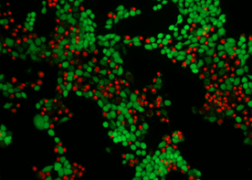
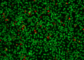 Figure2:differert cells were treated with 5μM Camptothecin for 4 h and photographed.


CCK-8
Cell Counting Kit-8(CCK-8) is a rapid and highly sensitive kit based on WST-8, which is widely used in the detection of cell activity and cytotoxicity.
Wst-8 is a compound similar to MTT. In the presence of electron carrier, WST-8 can be reduced by dehydrogenase in mitochondria to form water-soluble orange-yellow dark (Formazan) products, and the amount of Formazan produced is proportional to the number of viable cells. The more and faster the cell proliferation, the darker the color is. The amount of viable cells can be calculated indirectly by measuring the absorbance at 450 nm.
Because the CCK-8 solution is very stable and has little cytotoxicity, longer incubation periods, such as 24 to 48 hours, can be performed. The detection sensitivity of the CCK-8 kit is higher than that of any other tetrazolium salt, such as MTT, XTT or MTS.

|
High sensitivity |

|
Wide linear and perfect reproducibility |

|
Low toxicity, simple operation |

|
Multiple applications, drug screening, tumor drug sensitivity etc. |
| Product Name | Cat. No | Size |
|---|---|---|
| Enhanced Cell Counting Kit 8 (WST-8/CCK8) | E-CK-A362 | 100/500/10000 T |
![Enhanced Cell Counting Kit 8 (WST-8 / CCK8)[362]](https://www.elabscience.com/Uploads/Editor/2021-03-04/60408da4b7ee8.png)
Experiment Guide
LDH
Lactate dehydrogenase (LDH) is a kind of oxidoreductase that only exist in cytoplasm of all kinds of cells, it can be released into supernatant during the process of cell death or damage. The quantitation of cytotoxicity can be measured by measuring the extracellular LDH activity through enzymetic reaction.
LDH catalyzes the reaction of lactic acid with NAD+ to produce pyruvic acid and NADH. Under the action of PMS, electrons are transfered from NADH to WST-8 to produce the yellow formazan dye which has a characteristic absorption peak at 450 nm.
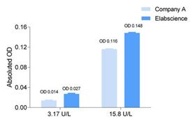
- Simple:Positively correlated with the cell death rate (With greater cytotoxicity, comes greater optical density)
- Higher Sensitivity:Giving higher OD value for the same sample (Comparing with other LDH cytotoxicity assay kits)
- Faster:Much shorter reaction time (As short as 10 min)
- Widely Application:A more general application with micro-plate reader using 450 nm detection
| Product Name | Cat. No | Size |
|---|---|---|
| Lactate Dehydrogenase (LDH) Cytotoxicity Colorimetric Assay Kit | E-BC-K771-M | 96 T |
Lactate Dehtdrogenase (LDH) Cytotoxicity Colorimetric Assay Kit
Sample Type: Cell
-
 Higher Sensitivity for the same sample (Comparing with other LDH cytotoxicity assay kits)
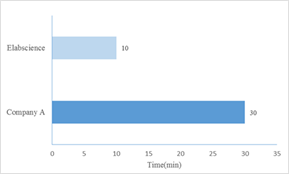 Much shorter reaction time (As short as 10 min)