How To Choose Flow Cytometry Antibodies
2024-07-26Author:adminpraise:0
Choosing high-quality and reliable fluorescent antibodies ensures the smooth running of flow cytometry and data analysis, especially multi-color flow cytometry that needs high resolution for color compensation. How to choose superior fluorescent antibodies for flow cytometry? Here are some key factors.
Three Key Factors
1. Commonly used clone numbers
There are usually several monoclonal antibodies with different clone numbers for a specific CD (clusters of differentiation) molecule. The more frequently the clone is used in flow cytometry, the more chance that you accomplish the experiments smoothly.
2. High SI (Staining Index)
High SI ensures good separation of the positive and negative cell populations, especially in experiments that need high resolution.
3. Low background binding
Though isotype antibodies or blocking reagents are usually used, sub-optimal conjugation of fluorescent dyes with antibodies significantly aggravate the background binding, especially with antibodies labeled by labeling kit without purification. Low background binding makes it much easier to determine the positive cell population from the negative one.
Why choose Elabscience@ flow cytometry antibodies?
Elabscience has selected commonly used clones as antibody source for flow cytometry
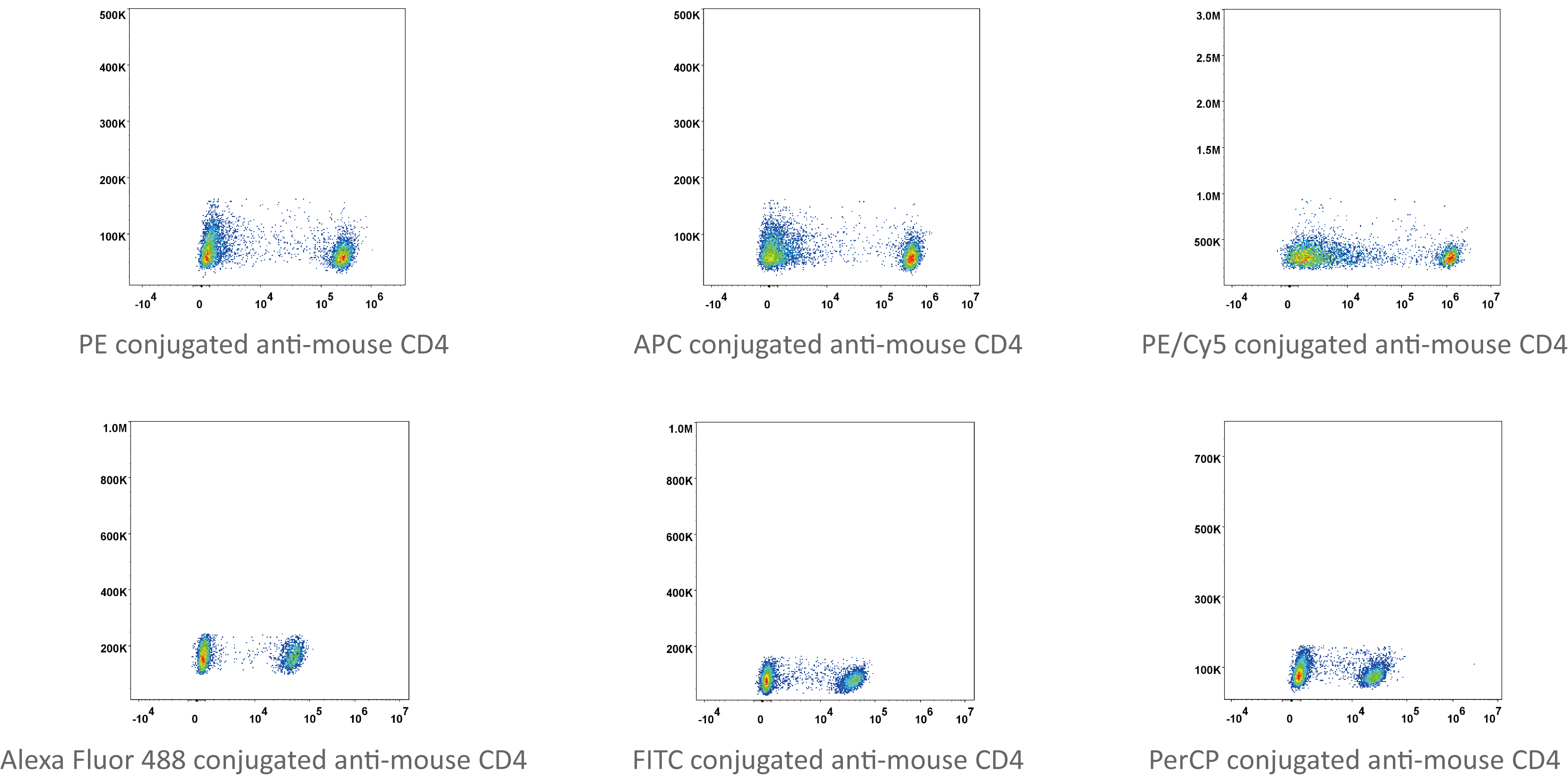
1. We have antibodies of six colors now and many more are on the way.

2. Elabscience@ flow cytometry antibodies have considerable SI (stainging index)
|
Fluorescent Dye |
Elabscience |
Manufacturer A |
|
PE anti-mouse CD4 |
219 |
158 |
|
APC anti-mouse CD4 |
238 |
200 |
|
Alexa Fluor@ 488 anti-mouse CD4 |
66 |
92 |
|
FITC anti-mouse CD4 |
61 |
56 |
|
PerCP anti-mouse CD4 |
31 |
9 |
Table 1. Comparison of SI of Elabscience@ flow cytometry antibodies with other manufacturers.
3. Elabscience@ flow cytometry antibodies have low background binding
With optimized conjugation technology, our flow cytometry antibodies can work with unstained cells instead of negative isotype control. Human blood lymphocytes stained with PE conjugated anti-human CD8a antibody. The negative population (empty blue curve) has nearly the same fluorescence level of the unstained cells (filled red curve).