Introduction of cell death--Cell Necrosis
2024-06-11Author:adminpraise:0
As the basic unit of an organism, cells live and die, just like humans. The "birth, aging, disease and death" of cells are closely related to human health. Cell death is necessary to maintain the function and morphology of tissues. The previous article introduced apoptosis and autophagy, and this article mainly introduces the related content of cell necrosis.
Definition of cell necrosis
Cell Necrosis is the cell death induced by extreme physical, chemical or other serious pathological factors, which is pathological cell death.
Cell itself, there is a "program", if the cells to work according to the "program" set run until death, so we'll call it programmed death is death. But if a cell has a little accident in the middle, out of the "programmed" control, then the cell that dies is non-programmed death, that is, cell necrosis.
Cell necrosis has long been considered as a passive death caused by pathology. However, recent studies have shown that cell necrosis may be another form of "programmed cell death" with important physiological functions, including the initiation of inflammatory response. Necrosis is adopted as an "alternate" mode of apoptosis when apoptosis cannot occur properly and cells must die.
Pathological changes in cell necrosis
Nuclear changes: this is the main morphological sign of cell necrosis, showing nuclear condensation, nuclear fragmentation, nuclear lysis;
The cytoplasm of change: Because the cytoplasmic solidification or dissolve, HE dyed dark red granule, such as liver cell necrosis of eosinophilic body;
Changes in the stroma: Due to the action of various lytic enzymes, the matrix disintegrates, the collagen fibers swell, break or liquefy, fuse with necrotic cells into a piece, and appear as red-stained granular unstructured material.
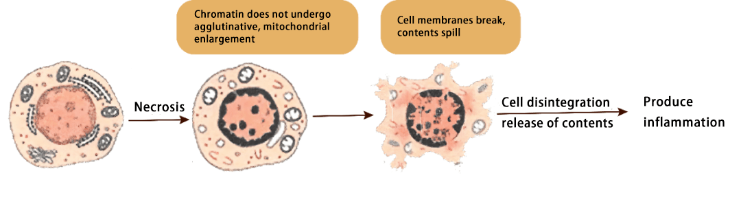
Figure 1. Schematic diagram of the process of cell necrosis
Types of cell necrosis
Cell necrosis can be classified into the following types based on protein degeneration in tissues:
1. Coagulation necrosis: Coagulation necrosis refers to the dry state of local tissue cells after death, also known as ischemic necrosis, cells due to water loss, protein coagulation, and the formation of dry coagulation. There are also some special types of coagulation necrosis, such as caseous necrosis, often caused by tuberculosis bacillus infection, due to the role of tuberculosis bacilli, resulting in tissue necrosis after yellow, soft caseous state;
2. Liquefaction necrosis: it is a state of liquefaction after cell death, called liquefaction necrosis, which is more common in tissues with less protein, more fat or water. It is easy to dissolve and liquidate after necrosis. Fat necrosis is also a kind of liquefaction necrosis, which is mainly liquid due to the disintegration and necrosis of fat cells.
3. Fibrinoid necrosis: this kind of necrosis can only be seen under the medical microscope, which is manifested as small strips or small lumps of unstructured tissue after necrosis, like fibrin, so it is called fibrinoid necrosis;
4. Gangrene: a secondary bacterial infection of necrotic tissue cells, resulting in the black and dark green appearance of these necrotic tissues. Gangrene includes dry gangrene, which is coagulation necrosis, and wet gangrene, which is similar to liquefaction necrosis. Some gangrene will produce a large amount of gas, called gas gangrene, this gas gangrene is often formed on the basis of liquefaction necrosis, belonging to the type of special wet gangrene.
Differentiation of necrosis from apoptosis and autophagy
Although the final results of apoptosis, autophagy and necrosis are very similar, their processes and manifestations are very different.
As a type of active end of life of cells, apoptosis plays a very important role in the process of maintaining the stability of the body. Its occurrence is caused by the gradual activation of the apoptotic pathway under the control of the program and does not produce inflammation. Autophagy is the phagocytosis of damaged organelles or aging proteins by intracellular lysosomes, which is a kind of self-protection of cells and usually triggers inflammation. Cell necrosis is often the type of acute end of life of cells, and cell contents will be released to the outside of the cell, leading to inflammation. They are all modes of cell death, but there are great differences in the causes, mechanisms, and morphology of each part of the cell
Table 1. Differences between Apoptosis, autophagy, and necrosis
|
|
Apoptosis |
Autophagy |
Necrosis |
|
Cause |
Induced by physiological or minor pathological stimuli |
Nutritional deficiency or hormone induction |
Pathological stimulator induced or severe injury |
|
Cytomembrane |
Membrane structure intact |
Membrane structure intact |
Membranolysis |
|
Changes in cell morphology |
Decrease, pyknosis |
Produce cavitation |
Cell swelling, enlargement, deformation |
|
DNA |
Degraded to 180~200 bp fragments |
Random degradation |
Random degradation |
|
Organelle |
Keep intact |
Phagocytosed by autophagosomes and eventually digested by lysosomes |
Deformation or swelling |
|
Lysosome |
Keep intact |
Forming autophagic lysosomes in the later stage |
Lysosomal destruction, enzyme spillage |
|
Results |
Formation of apoptotic bodies, phagocytosis by macrophages |
Cytoplasmic vacuolation, autophagosome formation, clearance of substances by lysosomes |
Cell rupture, lysis, phagocytosis by macrophages |
|
Inflammatory response |
Does not cause inflammation |
Can cause inflammation, which increases the risk of intestinal disease |
Cause phagocytosis of surrounding tissues by macrophages |
|
Mechanism |
Related to the protease Caspase gene family |
Occur under the action of lysosomes |
Relate to the expression of protein kinase RIP3 |
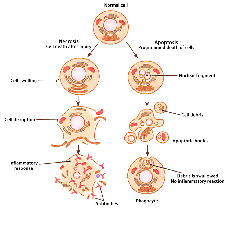
Figure 2. Comparison of Apoptosis and necrosis
How to detect cell necrosis?
The detection of cell necrosis first needs to determine whether the cell membrane is broken. The detection methods include:
1. Morphological observation: under transmission electron microscope or scanning electron microscope, if there is a body with intact membrane, it is apoptosis; If the membrane breaks, it is necrosis.
2. Immunofluorescence or flow method: PI or 7-AAD staining, because PI and 7-AAD can bind to DNA, if the cell membrane is broken, the dye can enter the cell and fluorescence.
Then we can determine whether it is programmed necrosis or passive necrosis, if it is a physical factor, such as an extreme environment, it is passive necrosis.
Conclusion
In the early study of cell death, Apoptosis and Necrosis were initially considered to be the two main modes of cell death. Apoptosis is a tightly regulated programmed active death mode, which does not release inflammatory factors. Necrosis, on the other hand, is an unexpected, unregulated, non-programmed passive mode of death that releases inflammatory factors. However, with the progress of research, more and more studies have shown that necrosis is also closely regulated by signaling pathways, called regulatory necrosis. The known regulatory necroptosis includes necroptosis, Ferroptosis, copper death, disulfoxide death, Parthanatos and cyclophilin D (CypD) -dependent necrosis.