Introduction of cell death--Apoptosis
2024-04-10Author:adminpraise:0
Cells in the human body are constantly renewed, and during this process the original cells will gradually die. This death includes natural aging and other non-aging pathways. There are various ways of cell death, including apoptosis, autophagy, pyroptosis, copper death, ferroptosis, etc.
Apoptosis
Apoptosis, also known as programmed cell death or cell suicide, is a process of active cell death under genetic control. Unlike necrosis, apoptosis does not release cellular contents into the surrounding environment.
Apoptosis exists widely in the biological world and occurs under normal physiological and pathological conditions. It plays a key role in embryonic development, tissue stability, body defense and immune response, and has therefore become one of the hot spots in biomedical research.
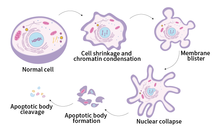
Figure 1. Apoptosis process
Characteristics of apoptosis
During the process of apoptosis, various characteristic physiological phenomena emerge. As the degree of apoptosis progresses further, distinct physiological manifestations also arise.
Early apoptosis stage: changes in cell membrane structure, extraversion of cell membrane phosphatidylserine (PS), activation of apoptosis-related proteins such as Bcl-2, activation of intracellular Caspase enzymes, collapse of mitochondrial membrane potential, etc.
Mid-stage of apoptosis: The cell population in the sub-G1 phase increases.
Late withering stage: cell nucleus shrinks, DNA fragments, cell membrane buds, and apoptotic bodies are formed.
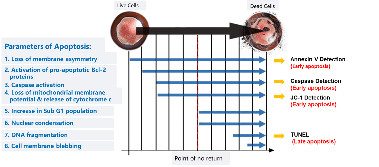
Figure 2. Schematic diagram of the characteristics of the apoptosis process.
Common detection methods of apoptosis
Based on the different physiological changes produced during cell apoptosis, different detection methods can be used to detect cell apoptosis.
01 Microscope observation
The morphological changes of apoptosis can be observed under a microscope, which can be observed through HE staining, acridine orange staining, and trypan blue staining through light microscopy or fluorescence microscopy, and cell apoptosis can be detected through color or nuclear and cytoplasmic status.
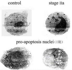
Figure 3. Electron microscopy observation of morphological changes in nuclear chromatin during Jurkat cell apoptosis (Source Network)
02 Annexin V detects cell membrane PS eversion
When cells undergo early apoptosis, PS on the cell membrane is everted. Annexin V is a protein that binds to PS. When cells undergo apoptosis, Annexin V connected to a fluorescent dye can be used to combine with the everted PS, and the occurrence of apoptosis can be determined by detecting the signal of the fluorescent group.
03 Detection of proteins activated during apoptosis
After the cell membrane loses symmetry, certain apoptosis-related proteins in the cell are activated, such as Bcl-2, BAX, FASL/TNFSF6, TNF-α, etc. The activated proteins in the cell can be determined by ELISA and other methods. Whether the cells are in a state of apoptosis.
04 Caspase enzyme activity detection
In the early stages of apoptosis, Caspase present in the cytoplasm is activated, triggering a series of apoptosis-related cascade reactions. Cell apoptosis can be determined by detecting the activity of Caspase.
05 JC-1 detects changes in mitochondrial membrane potential
The mitochondrial membrane potential of normal cells is high, and JC-1 can aggregate and emit red fluorescence; when cells undergo apoptosis, the mitochondrial membrane potential decreases and JC-1 cannot aggregate, so it exists as a green monomer. The occurrence of apoptosis can be judged by detecting changes in mitochondrial membrane potential.
06 TUNEL detects DNA fragmentation
In the late stage of apoptosis, DNA fragmentation occurs, and the exposed 3’-OH ends of fragmented DNA can be catalyzed by terminal deoxynucleotidyl transferase (TdT) to bind with fluorescently labeled dUTP. The fragmentation of DNA can be detected using a TUNEL assay kit to determine the occurrence of apoptosis in cells.
Elabscience® apoptosis detection reagents